|
|
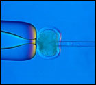
Enucleation of an oocyte |
|
|
Enucleation of an oocyte |
October 11, 2010
GERON (http): INTROD. START; STEM CELLS; OLIGODENDROCYTE; FINIS
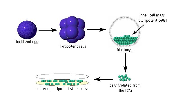
EMBRYONIC STEM CELLS
|
|
CLONING EMBRYONIC STEM CELLS
|
|
Figure 1. Comparison of Normal Development with Development during Reproductive Cloning and Therapeutic Cloning.
ADULT STEM CELLS
|
|
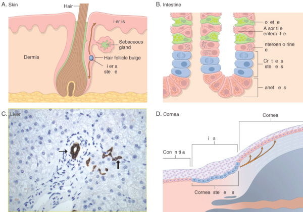
Figure 3-5
Stem-cell niches in various tissues. A,
Epidermal stem cells located in the bulge area of the hair follicle serve as a
stem cells for the hair follicle and the epidermis. B, Intestinal stem
cells are located at the base of a colon crypt, above Paneth cells. C,
Liver stem cells (commonly known as oval cells) are located in the canals of
Hering (thick arrow), structures that connect bile ductules (thin
arrow) with parenchymal hepatocytes (bile duct and Hering canals are stained
for cytokeratin 7; D, Corneal stem cells are located in the limbus
region, between the conjunctiva and the cornea.
courtesy of Tania Roskams, M.D., University of Leuven).
(Courtesy of T-T Sun, New York University, New York, NY.)
Kumar: Robbins and
Cotran: Pathologic Basis of Disease,
Table 1. Adult Human Stem Cells and Their Primary Direction of Differentiation.
|
Cell Type |
Tissue-Specific Location |
Cells or Tissues Produced |
|
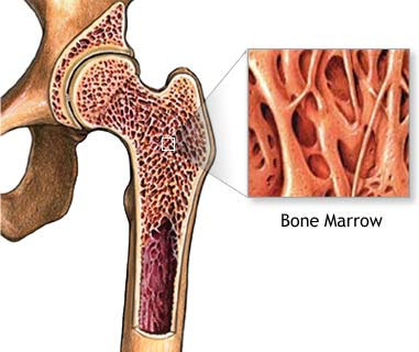
Hematopoietic |
Bone marrow, peripheral blood |
Bone marrow and blood lymphohematopoetic cells |
|
Mesenchymal stem cell |
Bone marrow, peripheral blood |
Bone, cartilage, tendon, adipose tissue, muscle, marrow stroma, neural cells |
|
Neural stem cells |
Ependymal cells, astrocytes (subventricular zone) of the CNS |
Neurons, astrocytes, oligodendrocytes |
|
Hepatic stem cells |
In or near the terminal bile ductules (canals of Hering) |
Oval cells that subsequently generate hepatocytes and ductular cells |
|
Pancreatic Stem cells |
Intra-islet Nestin positive cells, oval cells, duct cells |
Beta cells |
|
Skeletal muscle Stem cells |
Muscle fibers |
Skeletal muscle fibers |
|
Stem cells of the skin (keratinocytes) |
Basal layer of the epidermis, bulge zone of the hair follicles |
Epidermis, hair follicles |
|
Epithelial stem cells of the lung |
Tracheal basal and mucus-secreting cells, bronciolar Clara cells, alveolar Type II pneumocyte |
Mucous and ciliated cells, type I and II pneumocytes |
|
Stem cells of the intestinal epithelium |
Epithelial cells located around the base of each crypt |
Paneth’s cells, brush-border enterocytes, mucus secreting goblet cells, enteroendocrine cells |
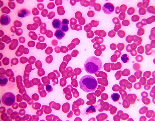
BLOOD CELLS
|
|
|
|
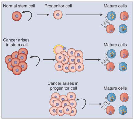
CANCERS and STEM CELLS
|
|
Figure 8-2
Target cells for neoplastic transformation. In many tissues in which cancers
arise, the stem cells are the only long-lived cells and are the only cells
capable of self-renewal. Because they are already are capable of extensive
self-renewal, they are good targets for neoplastic transformation. Dysregulation
of the self-renewal process may be simpler in these cells than in progenitor
cells that lack this ability. In order for progenitor cells to undergo malignant
transformation, they must acquire the ability to undergo extensive self-renewal
as a result of oncogenic mutations.
Abeloff:
Clinical Oncology,
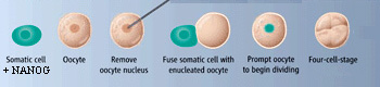
ALTERED
NUCLEAR
TRANSFER-
OOCYTE
ASSISTED
REPROGRAMMING
Somatic cell nuclear transfer of nucleus capable ONLY of becoming pleuripotent cells - NOT a human embryo
|
|
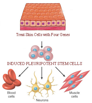
INDUCED PLEURIPOTENT STEM CELLS
|
|
 J
J