|
Virgin and Child, Enthroned, The Master of Moulins, 1499 |
FETAL
|
|
|
Virgin and Child, Enthroned, The Master of Moulins, 1499 |
FETAL
|
|
When sperm is deposited in the vagina, it travels through the cervix and into the Fallopian tubes. Conception usually takes place in the Fallopian tube. A single sperm penetrates the mother's egg cell, and the resulting cell is called a zygote.
The zygote contains all of the genetic information (DNA) necessary to become a child. Half of the genetic information comes from the mother’s egg, and half from the father’s sperm.
The zygote spends the next few days traveling down the Fallopian tube and divides to form a ball of cells. Further cell division creates an inner group of cells with an outer shell. This stage is called a "blastocyst". The inner group of cells will become the embryo, while the outer group of cells will become the membranes that nourish and protect it.
The blastocyst reaches the uterus at roughly the fifth day, and implants into the uterine wall on about day six. At this point in the mother's menstrual cycle, the endometrium (lining of the uterus) has grown and is ready to support a fetus. The blastocyst adheres tightly to the endometrium, where it receives nourishment via the mother's bloodstream.
The cells of the embryo now multiply and begin to take on specific functions. This process is called differentiation, which produces the varied cell types that make up a human being (such as blood cells, kidney cells, and nerve cells).
There is rapid growth, and the baby's main external features begin to take form. It is during this critical period of differentiation (most of the first trimester) that the growing baby is most susceptible to damage from:
Alcohol, certain prescription and recreational drugs, and other substances that cause birth defects
Infection (such as rubella or cytomegalovirus)
Radiation from x-rays or radiation therapy
Nutritional deficiencies
The following list describes specific changes by week.
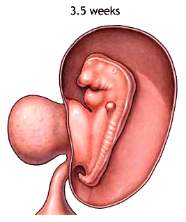
Week 3
beginning development of the brain, spinal cord, and heart. Heart begins to beat at end of third week.
beginning development of the gastrointestinal tract
 |
Weeks 4 to 5
formation of tissue that develops into the vertebra and some other bones
further development of the heart which now beats at a regular rhythm
movement of rudimentary blood through the main vessels
beginning of the structures of the eye and ears
the brain develops into five areas and some cranial nerves are visible
arm and leg buds are visible
Week 6
beginning of formation of the lungs
further development of the brain
arms and legs have lengthened with foot and hand areas distinguishable
hands and feet have digits, but may still be webbed
Week 7
nipples and hair follicles form
elbows and toes visible
all essential organs have at least begun to form
 |
Week 8
rotation of intestines
facial features continue to develop
the eyelids are more developed
the external features of the ear begin to take their final shape
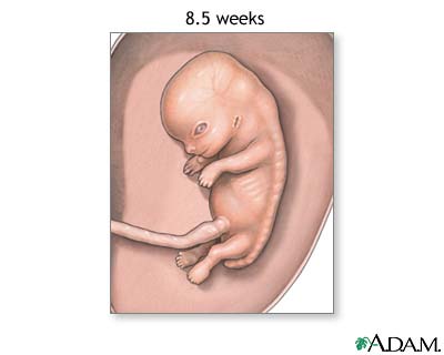 |
The end of the eighth week marks the end of the "embryonic period" and the beginning of the "fetal period".
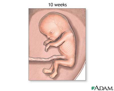 |
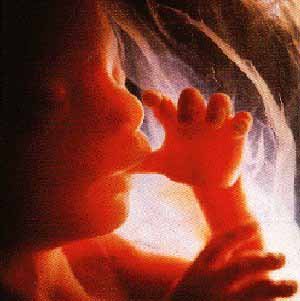
Weeks 9 to 12
the fetus reaches a length of 3.2 inches
the head comprises nearly half of the fetus' size
the face is well formed
eyelids close and will not reopen until about the 28th week
tooth buds appear for the baby teeth
limbs are long and thin
the fetus can make a fist with its fingers
genitals appear well differentiated
red blood cells are produced in the liver
 |
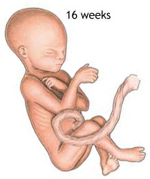
Weeks 13 to 16
the fetus reaches a length of about 6 inches
a fine hair develops on the head called lanugo
fetal skin is almost transparent
more muscle tissue and bones have developed, and the bones become harder
the fetus makes active movements
sucking motions are made with the mouth
meconium is made in the intestinal tract
the liver and pancreas produce their appropriate fluid secretions
 |
Week 20
the fetus reaches a length of 8 inches
lanugo hair covers entire body
eyebrows and lashes appear
nails appear on fingers and toes
the fetus is more active with increased muscle development
"quickening" usually occurs (the mother can feel the fetus moving)
fetal heartbeat can be heard with a stethoscope
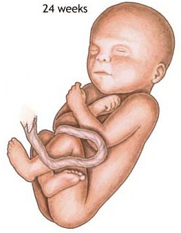 |
Week 24
the fetus reaches a length of 11.2 inches
the fetus weighs about 1 lb. 10 oz.
eyebrows and eyelashes are well formed
all the eye components are developed
the fetus has a hand and startle reflex
footprints and fingerprints forming
alveoli (air sacs) forming in lungs
 |
Weeks 25 to 28
the fetus reaches a length of 15 inches
the fetus weighs about 2 lbs. 11 oz.
rapid brain development
nervous system developed enough to control some body functions
eyelids open and close
respiratory system, while immature, has developed to the point where gas exchange is possible
a baby born at this time may survive, but the possibilities for complications and death remain high
STATISTICS
on VIABILITY
from
Ford, The Prenatal
Person, ch. 9, "Newborns"
Table 9.3: Neonatal intensive care unit, Loyola University Medical Center, survival rates, 1990—4
|
Gestational age, weeks |
Survivors % |
|
22—3 |
19 % |
|
24—5 |
63 % |
|
26—7 |
88 % |
Source: modified from “Neonatal Survival Rates” in Cambridge Quarterly of Healthcare Ethics 8 (1999):162.
Table 9.4: Survival rates by gestational age, 1994–6
|
Gestational age (weeks) |
Live births |
Survivors percentage |
|
23 |
31 |
35.5 % |
|
24 |
36 |
65.6 % |
|
25 |
46 |
73.9 % |
|
26 |
64 |
85.9 % |
|
28 |
75 |
96.0 % |
|
32 |
262 |
98.1 % |
Source: modified and calculated from data in the Royal Women’s Hospital in Melbourne, Medical Journal of Australia, June 7, 1999.
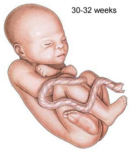 |
Weeks 29 to 32
the fetus reaches a length of about 15-17 inches
the fetus weighs about 4 lbs. 6 oz.
rapid increase in the amount of body fat
rhythmic breathing movements occur, but lungs are not fully mature
bones are fully developed, but still soft and pliable
fetus begins storing iron, calcium, and phosphorus
Week 36
the fetus reaches a length of about 16-19 inches
the fetus weighs about 5 lbs. 12 oz. to 6 lbs. 12 oz.
lanugo begins to disappear
increase in body fat
fingernails reach the end of the fingertips
a baby born at 36 weeks has a high chance of survival, but may require some medical interventions
Weeks 37 to 40
considered full-term at 37 weeks
may be 19 to 21 inches in length
lanugo is gone except for on the upper arms and shoulders
fingernails extend beyond fingertips
small breast buds are present on both sexes
head hair is now coarse and thicker