|
|
Virgin and Child, Master of Moulins, 1499 |
|
|
|
Virgin and Child, Master of Moulins, 1499 |
|
1. EMBRYONIC DEVELOPMENT; 2. FETAL DEVELOPMENT; 3. VIABILITY
PLEASE note that the black-and-white photographs shown here were assembled by the Carnegie Embryology Institute over several decades, beginning in 1914, as part of a national effort to identify, describe, and standardize the stages of human embryological and fetal development. The photographs are of embryos that were the result of spontaneous miscarriages or that were discovered during a necessary hysterectomy. In other words, these are not photographs of intentionally-aborted embryos.
For embryo sizes compared with a US penny:
http://virtualhumanembryo.lsuhsc.edu/HEIRLOOM/Stages/Stages_intro.html
|
|
|
|
|
|
|
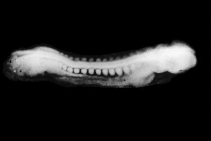
WEEK 3 |
|
|
|
|
|
|
|
|
STAGE
11; |
|
|
|
|
|
|
|
|
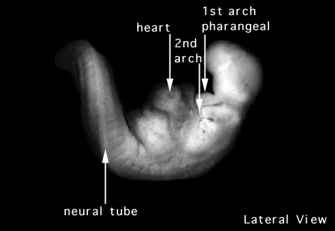
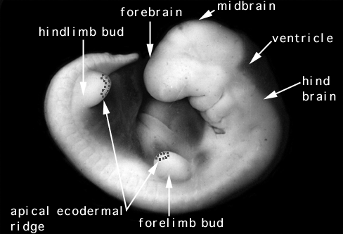
WEEK 4 |
|
|
|
|
|
|
|
|
STAGE
12; |
|
|
|
|
|
|
|
|
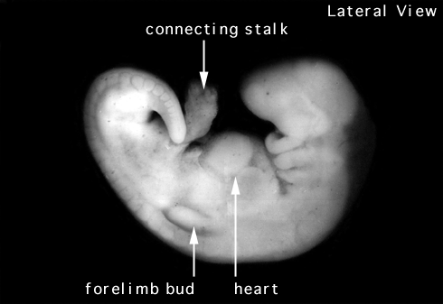
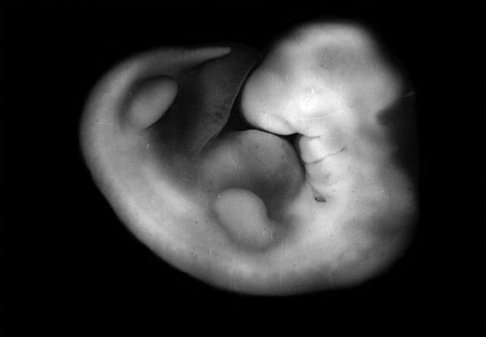
WEEK 5 |
|
|
|
|
|
|
|
|
STAGE
13; |
|
|
STAGE
14; |
|
|
STAGE
15; |
|
|
|
|
|
|
|
|
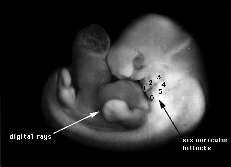
WEEK 6 |
|
|
|
|
|
|
|
|
STAGE
16; |
|
|
STAGE
17; |
|
|
|
|
|
|
|
|
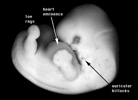
WEEK 7 |
|
|
|
|
|
|
|
|
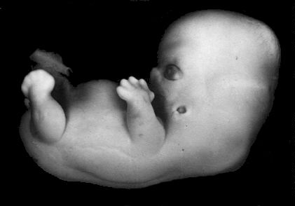
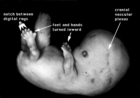
STAGE
18; |
|
|
STAGE
19; |
|
|
STAGE
20; |
|
|
STAGE
21; |
|
|
|
|
|
|
|
|
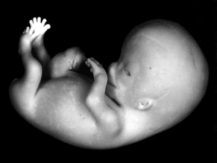
WEEK 8 |
|
|
|
|
|
|
|
|
STAGE
22; |
|
|
STAGE
23; |
|
|
|
|
|
|
|
The end of the eighth week marks the END of the “EMBRYONIC PERIOD” and the BEGINNING of the “FETAL PERIOD”.
WEEKS 9-12
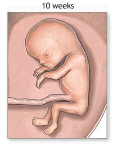 |
the fetus reaches a length of 3.2 inches
the head comprises nearly half of the fetus' size
the face is well formed
eyelids close and will not reopen until about the 28th week
tooth buds appear for the baby teeth
limbs are long and thin
the fetus can make a fist with its fingers
genitals appear well differentiated
red blood cells are produced in the liver
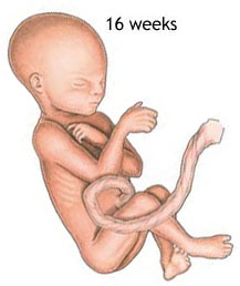
WEEKS 13-16

the fetus reaches a length of about 6 inches
a fine hair develops on the head called lanugo
fetal skin is almost transparent
more muscle tissue and bones have developed, and the bones become harder
the fetus makes active movements
sucking motions are made with the mouth
meconium is made in the intestinal tract
the liver and pancreas produce their appropriate fluid secretions
WEEK 20
 |
the fetus reaches a length of 8 inches
lanugo hair covers entire body
eyebrows and lashes appear
nails appear on fingers and toes
the fetus is more active with increased muscle development
"quickening" usually occurs (the mother can feel the fetus moving)
fetal heartbeat can be heard with a stethoscope
STATISTICS
on VIABILITY
from
Ford, The Prenatal
Person, ch. 9, "Newborns"
Table 9.3: Neonatal intensive care unit, Loyola University Medical Center, survival rates, 1990—4
|
Gestational age, weeks |
Survivors % |
|
22—3 |
19 % |
|
24—5 |
63 % |
|
26—7 |
88 % |
Source: modified from “Neonatal Survival Rates” in Cambridge Quarterly of Healthcare Ethics 8 (1999):162.
Table 9.4: Survival rates by gestational age, 1994–6
|
Gestational age, weeks |
Survivors percentage |
Live births |
|
23 |
35.5 % |
31 |
|
24 |
65.6 % |
36 |
|
25 |
73.9 % |
46 |
|
26 |
85.9 % |
64 |
|
28 |
96.0 % |
75 |
|
32 |
98.1 % |
262 |
Source: modified and calculated from data in the Royal Women’s Hospital in Melbourne, Medical Journal of Australia, June 7, 1999.
WEEK 24
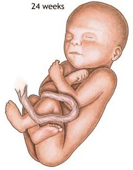 |
the fetus reaches a length of 11.2 inches
the fetus weighs about 1 lb. 10 oz.
eyebrows and eyelashes are well formed
all the eye components are developed
the fetus has a hand and startle reflex
footprints and fingerprints forming
alveoli (air sacs) forming in lungs
WEEKS 25-28

the fetus reaches a length of 15 inches
the fetus weighs about 2 lbs. 11 oz.
rapid brain development
nervous system developed enough to control some body functions
eyelids open and close
respiratory system, while immature, has developed to the point where gas exchange is possible
a baby born at this time may survive, but the possibilities for complications and death remain high
WEEKS 29-32
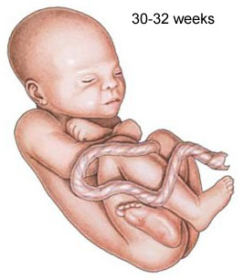 |
the fetus reaches a length of about 15-17 inches
the fetus weighs about 4 lbs. 6 oz.
rapid increase in the amount of body fat
rhythmic breathing movements occur, but lungs are not fully mature
bones are fully developed, but still soft and pliable
fetus begins storing iron, calcium, and phosphorus
WEEK 36
the fetus reaches a length of about 16-19 inches
the fetus weighs about 5 lbs. 12 oz. to 6 lbs. 12 oz.
lanugo begins to disappear
increase in body fat
fingernails reach the end of the fingertips
a baby born at 36 weeks has a high chance of survival, but may require some medical interventions
WEEKS 37-40
considered full-term at 37 weeks
may be 19 to 21 inches in length
lanugo is gone except for on the upper arms and shoulders
fingernails extend beyond fingertips
small breast buds are present on both sexes
head hair is now coarse and thicker
This Webpage was created for a workshop held at Saint Andrew's Abbey, Valyermo, California in 2003