|
|
|
|
|
|
Published at www.nejm.org February 3, 2010 (10.1056/NEJMoa0905370)
Martin M. Monti, Ph.D., Audrey Vanhaudenhuyse, M.Sc., Martin R. Coleman, Ph.D., Melanie Boly, M.D., John D. Pickard, F.R.C.S., F.Med.Sci., Luaba Tshibanda, M.D., Adrian M. Owen, Ph.D., and Steven Laureys, M.D., Ph.D.
ABSTRACT
Background The differential diagnosis of disorders of consciousness is challenging. The rate of misdiagnosis is approximately 40%, and new methods are required to complement bedside testing, particularly if the patient’s capacity to show behavioral signs of awareness is diminished.
Methods At two major referral centers in Cambridge, United Kingdom, and Liege, Belgium, we performed a study involving 54 patients with disorders of consciousness. We used functional magnetic resonance imaging (MRI) to assess each patient’s ability to generate willful, neuroanatomically specific, blood-oxygenation-level–dependent responses during two established mental-imagery tasks. A technique was then developed to determine whether such tasks could be used to communicate yes-or-no answers to simple questions.
Results Of the 54 patients enrolled in the study, 5 were able to willfully modulate their brain activity. In three of these patients, additional bedside testing revealed some sign of awareness, but in the other two patients, no voluntary behavior could be detected by means of clinical assessment. One patient was able to use our technique to answer yes or no to questions during functional MRI; however, it remained impossible to establish any form of communication at the bedside.
Conclusions These results show that a small proportion of patients in a vegetative or minimally conscious state have brain activation reflecting some awareness and cognition. Careful clinical examination will result in reclassification of the state of consciousness in some of these patients. This technique may be useful in establishing basic communication with patients who appear to be unresponsive.
In recent years, improvements in intensive care have led to an increase in the number of patients who survive severe brain injury. Although some of these patients go on to have a good recovery, others awaken from the acute comatose state but do not show any signs of awareness. If repeated examinations yield no evidence of a sustained, reproducible, purposeful, or voluntary behavioral response to visual, auditory, tactile, or noxious stimuli, a diagnosis of a vegetative state — or “wakefulness without awareness” — is made.1,2,3,4,5 Some patients remain in a vegetative state permanently. Others eventually show inconsistent but reproducible signs of awareness, including the ability to follow commands, but they remain unable to communicate interactively. In 2002, the Aspen Neurobehavioral Conference Work Group coined the term “minimally conscious state” to describe the condition of such patients, thereby adding a new clinical entity to the spectrum of disorders of consciousness.6
There are two main goals in the clinical assessment of patients in a vegetative or minimally conscious state. The first goal is to determine whether the patient retains the capacity for a purposeful response to stimulation, however inconsistent. Such a capacity, which suggests at least partial awareness, distinguishes minimally conscious patients from those in a vegetative state and therefore has implications for subsequent care and rehabilitation, as well as for legal and ethical decision making. Unfortunately, the behavior elicited from these patients is often ambiguous, inconsistent, and constrained by varying degrees of paresis, making it very challenging to distinguish purely reflexive from voluntary behaviors. Nevertheless, in the absence of an absolute measure, awareness has to be inferred from a patient’s motor responsiveness; this fact undoubtedly contributes to the high rate of diagnostic errors (approximately 40%) in this group of patients.7,8,9
The second goal of clinical assessment is to harness and nurture any available response, through intervention, into a form of reproducible communication, however rudimentary. The acquisition of any interactive and functional verbal or nonverbal method of communication is an important milestone. Clinically, consistent and repeatable communication demarcates the upper boundary of a minimally conscious state.6
In this article, we present the results of a study conducted between November 2005 and January 2009 in which functional magnetic resonance imaging (MRI) was routinely used in the evaluation of a group of 54 patients with a clinical diagnosis of being in a vegetative state or a minimally conscious state. In light of a previous single-case study that showed intact awareness in a patient who met the clinical criteria for being in a vegetative state,10 our investigation had two main aims. The first aim was to determine what proportion of this group of patients could also reliably and repeatedly modulate their functional MRI responses, reflecting preserved awareness. The second aim was to develop and validate a method that would allow such patients to functionally communicate yes-or-no responses by modulating their own brain activity, without training and without the need for any motor response.
Methods
Patients
A convenience sample of 54 patients with severe brain injury, including 23 in a vegetative state and 31 in a minimally conscious state, underwent functional MRI as a means of evaluating their performance on motor and spatial imagery tasks. Characteristics of the patients are shown in Table 1, and the inclusion criteria are described in the Supplementary Appendix, available with the full text of this article at NEJM.org. Written informed consent was obtained from the legal guardians of all patients. The motor and spatial imagery tasks have been well validated in healthy control subjects10,11,12 and are known to be associated with distinct functional MRI activity in the supplementary motor area and the parahippocampal gyrus.
|
The method to detect functional communication was first tested for
feasibility and robustness in 16 healthy control subjects (9 men and
7 women) with no history of a neurologic disorder. Once validated,
the tasks were given to one patient (Patient 23 in
Table 1 and
Figure 1), who had received a diagnosis of being in a permanent
vegetative state 17 months after a traffic accident; this diagnosis
was confirmed by a month-long specialized assessment 3.5 years after
the injury. At the time of admission for functional MRI scanning (5
years after the ictus), the patient was assumed to remain in a
vegetative state, although extensive behavioral testing after the
functional MRI revealed reproducible, but inconsistent, responses
indicative of a minimally conscious state. (The
Supplementary Appendix includes detailed results and a
description of the clinical assessment of this patient.)
|
Imagery Tasks
While in the functional MRI scanner, all patients were asked to perform two imagery tasks. In the motor imagery task, they were instructed to imagine standing still on a tennis court and to swing an arm to “hit the ball” back and forth to an imagined instructor. In the spatial imagery task, participants were instructed to imagine navigating the streets of a familiar city or to imagine walking from room to room in their home and to visualize all that they would “see” if they were there. First, two so-called localizer scanning sessions were conducted in which the patients were instructed to alternate 30-second periods of mental imagery with 30-second periods of rest. Each scan included five rest–imagery cycles. The beginning of each imagery period was cued with the spoken word “tennis” or “navigation,” and rest periods were cued with the word “relax.”
Communication Task
After the localizer scans had been obtained, all 16 control subjects and 1 patient underwent functional MRI during which they attempted to answer questions by modulating their brain activity, and a set of so-called communication scans were obtained. Before each of these imaging sessions, participants were asked a yes-or-no question (e.g., “Do you have any brothers?”) and instructed to respond during the imaging session by using one type of mental imagery (either motor imagery or spatial imagery) for “yes” and the other for “no.” The nature of the questions ensured that the investigators would not know the correct answers before judging the functional MRI data. Participants were asked to respond by thinking of whichever imagery corresponded to the answer that they wanted to convey. Communication scanning was identical to localizer scanning with the exception that the same neutral word “answer” was used to cue each response to a question (with “relax” used as the cue for rest periods). Cues were delivered once, at the beginning of each period. Three communication scans (with one question per scan) were obtained for each of the 16 healthy control subjects. To maximize statistical power, six communication scans (with one question per scan) were obtained for the patient.
Statistical Analysis
Analyses were performed with the use of FSL software, version 4.1.13 Data analysis included standard functional MRI preprocessing steps (functional MRI acquisition and preprocessing are described in the Supplementary Appendix). For each scan, a general linear model contrasting periods of active imagery with periods of rest was computed. All contrasts were limited to the brain locations within the supplementary motor area and the parahippocampal gyrus, as defined in the Harvard–Oxford Cortical Structural Atlas (available in FSL software), and a threshold was established, with gaussian random-fields theory, at a cluster-level z value of more than 2.3 (corrected P<0.05). The defined regions of interest were transformed from standard space (according to the criteria of the Montreal Neurological Institute) to fit each subject’s structural image, with the use of a method involving 12 degrees of freedom.
To determine whether the imagery tasks produced the expected activations in predefined neuroanatomical locations, two scans were compared for each participant: motor imagery and spatial imagery. The multiple localizer scanning sessions of the patient who also underwent communication scanning were averaged with the use of a fixed-effects model.
Answers provided during the communication scanning were assessed with the use of a two-step procedure. First, activity in the two regions of interest (the supplementary motor area and the parahippocampal gyrus) identified during the localizer scanning was quantitatively characterized (with the use of the average generalized linear model estimate for each region of interest). Next, a similarity metric (described in the Supplementary Appendix) was computed to quantify how closely the activity in the regions of interest on each communication scan matched each localizer scan.
Results
Responses to the Imagery Tasks
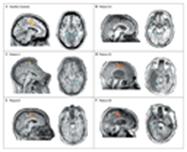
Among the 54 patients, we identified 5 who could willfully modulate their brain activity (Figure 1). In all five of these patients, the functional MRI scans associated with motor imagery, as compared with spatial imagery, showed considerable activation in the supplementary motor area. In four of the five patients, the scans associated with spatial imagery, as compared with motor imagery, showed activation in the parahippocampal gyrus. Furthermore, the time course of activity within the two regions of interest was sustained for 30 seconds and was associated with the delivery of the verbal cues (Figure 2). These results closely match the pattern observed in the healthy control subjects (Figure 1, and the Supplementary Appendix). Four of the five patients were considered to be in a vegetative state (including Patient 4, who has been described previously10), and all five patients had a traumatic brain injury (Table 1).
|
Responses to the Communication Task
Each of the 16 healthy control subjects underwent functional MRI to obtain three communication scans. For all 48 questions in the communication task, the correct answer was determined with 100% accuracy by comparing the activations shown on the communication scans with the activations shown on two localizer scans. In all subjects, the pattern produced in response to each question was quantitatively more similar to the pattern observed in the localizer scan for the imagery task that was associated with the factually correct answer; this answer was verified after the analysis. Figure 2B and 2D and Figure 3B and 3D show this similarity in a healthy control. In this subject, the activation associated with the imagery period as compared with the rest period for question 1 resulted in extensive activation in the supplementary motor area and minimal activity in the parahippocampal gyrus (Figure 4). This pattern was almost identical to that observed in the activation associated with the motor imagery period as compared with the rest period in the motor localizer scan. Conversely, the imagery period as compared with the rest period for questions 2 and 3 was associated with extensive activation of the parahippocampal gyrus and, to a lesser extent, the supplementary motor area; these findings closely matched the activation seen in the spatial localizer scan. Similar patterns were observed in 9 of 16 control subjects. In the remaining seven control subjects, the distinction between tasks was even clearer; thus, a double dissociation was observed between activity in the supplementary motor area for motor imagery and activity in the parahippocampal gyrus for spatial navigation (see the Supplementary Appendix).
|
|
To assess whether such an approach could be used in a patient with
impaired consciousness, one of the patients who had reliable
responses during the two imagery tasks (Patient 23) was also asked
six yes-or-no autobiographical questions and instructed to respond by
thinking of one type of imagery (either motor imagery or spatial
imagery) for an affirmative answer and the other type of imagery for
a negative answer.
In this patient, the activity observed on the communication scan in response to five of the six questions closely matched that observed on one of the localizer scans (Figure 2A and 2C and Figure 3A and 3C). For example, in response to the question “Is your father’s name Alexander?” the patient responded “yes” (correctly) with activity that matched that observed on the motor-imagery localizer scan (Figure 3A). In response to the question “Is your father’s name Thomas?” the patient responded “no” (also correctly) with activity that matched that observed in the spatial-imagery localizer scan (Figure 3C).
The relative-similarity analysis confirmed, quantitatively, that the activity observed on the communication scans accurately reproduced that observed on the localizer scans within the bounds of normal variability for five of the six questions (Figure 4, and Tables A1 and A2 in the Supplementary Appendix). In addition, for those same five questions, the pattern produced always matched the factually correct answer. Only one question, the last one, could not be decoded. However, this was not because the “incorrect” pattern of activation was observed, but rather because virtually no activity was observed within the regions of interest.
Discussion
In this study, functional MRI was used to determine the incidence of undetected awareness in a group of patients with severe brain injuries. Of the 54 patients, 5 with traumatic brain injuries were able to modulate their brain activity by generating voluntary, reliable, and repeatable blood-oxygenation-level–dependent responses in predefined neuroanatomical regions when prompted to perform imagery tasks. No such responses were observed in any of the patients with nontraumatic brain injuries. Four of the five patients who were able to generate these responses were admitted to the hospital with a diagnosis of being in a vegetative state. When these four patients were thoroughly retested at the bedside, some behavioral indicators of awareness could be detected in two of them. However, the other two patients remained behaviorally unresponsive at the bedside, even after the functional MRI results were known and despite repeated testing by a multidisciplinary team. Thus, in a minority of cases, patients who meet the behavioral criteria for a vegetative state have residual cognitive function and even conscious awareness.14,15
We conducted additional tests in one of the five patients with evidence of awareness on functional MRI, and we found that he had the ability to apply the imagery technique in order to answer simple yes-or-no questions accurately. Before the scanning was performed, the patient had undergone repeated evaluations indicating that he was in a vegetative state, including a month-long specialized assessment by a highly trained clinical team. At the time of scanning, however, thorough retesting at the bedside showed reproducible but highly fluctuating and inconsistent signs of awareness (see the Supplementary Appendix), findings that are consistent with the diagnosis of a minimally conscious state. Nonetheless, despite the best efforts of the clinical team, it was impossible to establish any functional communication at the bedside, and the results of the behavioral examination remained ambiguous and inconsistent. In contrast, the functional MRI approach allowed the patient to establish functional and interactive communication. Indeed, for five of the six questions, the patient had a reliable neural response and was able to provide the correct answer with 100% accuracy. For the remaining question — the last question of the imaging session — the lack of activity within the regions of interest precluded any analysis of the results. Whether the patient fell asleep during this question, did not hear it, simply elected not to answer it, or lost consciousness cannot be determined.
Although the functional MRI data provided clear evidence that the patient was aware and able to communicate, it is not known whether either ability was available during earlier evaluations. It is possible that he was in a vegetative state when the diagnosis was received at 17 months and again 3.5 years after injury and subsequently regained some aspects of cognitive functioning. Alternatively, the patient may have been aware during previous assessments but unable to produce the necessary motor response required to signal his state of consciousness. If this was the case, then the clinical diagnosis of a vegetative state was entirely accurate in the sense that no behavioral markers of awareness were evident. That said, the diagnosis did not accurately reflect the patient’s internal state of awareness and level of cognitive functioning at the time. Given that all previous assessments were based on behavioral observations alone, these two possibilities are indistinguishable.
Among 49 of the 54 patients included in this study, no significant functional MRI changes were observed during the imagery tasks. In these patients, it is not possible to determine whether the negative findings were the result of the low “sensitivity” of the method (e.g., failure to detect small effects), or whether they genuinely reflect the patients’ limited cognitive abilities. Some patients, for example, may have been unconscious (permanently or transiently) during scanning. Similarly, in some awake and aware patients who were in a minimally conscious state, the tasks may simply have exceeded their residual cognitive capabilities. Deficits in language comprehension, working memory, decision making, or executive function would have prevented successful completion of the imagery tasks. However, positive results, whether observed with or without corroborative behavioral data, do confirm that all such processes were intact and that the patient must have been aware.
In summary, the results of this study show the potential for functional MRI to bridge the dissociation that can occur between behavior that is readily observable during a standardized clinical assessment and the actual level of residual cognitive function after serious brain injury.14,15,16 Thus, among 23 patients who received a diagnosis of being in a vegetative state on admission, 4 were shown to be able to willfully modulate their brain activity through mental imagery; this fact is inconsistent with the behavioral diagnosis. In two of these patients, however, subsequent assessment at the bedside revealed some behavioral evidence of awareness, a finding that underscores the importance of thorough clinical examination for reducing the rate of misdiagnosis in such patients. Nonetheless, in the two remaining patients, no evidence of awareness could be detected at the bedside by an experienced clinical team, even after the results of the functional MRI examination were known. This finding indicates that, in some patients, motor function can be so impaired that bedside assessments based on the presence or absence of a behavioral response may not reveal awareness, regardless of how thoroughly and carefully they are administered. In patients without a behavioral response, it is clear that functional MRI complements existing diagnostic tools by providing a method for detecting covert signs of residual cognitive function17,18,19,20 and awareness.10
In addition, this study showed that in one patient with severe impairment of consciousness, functional MRI established the patient’s ability to communicate solely by modulating brain activity, whereas this ability could not be established at the bedside. In the future, this approach could be used to address important clinical questions. For example, patients could be asked if they are feeling any pain, and this information could be useful in determining whether analgesic agents should be administered. With further development, this technique could be used by some patients to express their thoughts, control their environment, and increase their quality of life.
Supported by grants from the Medical Research Council (U.1055.01.002.00007.01 and U.1055.01.002.00001.01), the European Commission (Disorders and Coherence of the Embodied Self, Mindbridge, Deployment of Brain–Computer Interfaces for the Detection of Consciousness in Nonresponsive Patients, and Consciousness: A Transdisciplinary, Integrated Approach), Fonds de la Recherche Scientifique, the James S. McDonnell Foundation, the Mind Science Foundation, the Reine Elisabeth Medical Foundation, the Belgian French-Speaking Community Concerted Research Action, University Hospital of Liege, the University of Liege, and the National Institute for Health Research Biomedical Research Centre (Neurosciences Theme).
No potential conflict of interest relevant to this article was reported.
We thank Daniel Gary Wakeman for his helpful discussions.
Source Information
From the Medical Research Council Cognition
and Brain Sciences Unit (M.M.M., A.M.O.), the Impaired Consciousness Study
Group, Wolfson Brain Imaging Centre, University of Cambridge (M.R.C.), and the
Division of Academic Neurosurgery, Addenbrooke’s Hospital (J.D.P.) — all in
Cambridge, United Kingdom; and the Coma Science Group, Cyclotron Research
Center, University of Liege (A.V., M.B., S.L.), and the Departments of Neurology
(S.L., M.B.) and Neuroradiology (L.T.), University Hospital of Liege, Liege; and
Fonds de la Recherche Scientifique, Brussels (A.V., S.L., M.B.) — all in
Belgium.
Dr. Monti and Ms. Vanhaudenhuyse contributed equally to this article.
This article (10.1056/NEJMoa0905370) was published on February 3, 2010, at
NEJM.org.
Address reprint requests to Dr. Owen at the Medical Research Council Cognition and Brain Sciences Unit, 15 Chaucer Rd., Cambridge CB2 7EF, United Kingdom, or at adrian.owen@mrc-cbu.cam.ac.uk.
References
This Webpage was created for a workshop held at Saint Andrew's Abbey, Valyermo, California in 1990