|
|
POTENTIAL
OPTIONS
Giotto, |
|
|
POTENTIAL
OPTIONS
Giotto, |
Rogerio A. Lobo, M.D.
The New England Journal of Medicine
July 7, 2005 Volume
353:64-73, Number 1
The choice of delaying pregnancy has become the norm for many women in developed countries. Among some women, however, achieving pregnancy may be difficult or impossible at a later time. The ability to preserve fertility with various methods has become a key issue for some women. Although the need is most pressing among women with cancer, the same therapeutic options may be available for many other women who are reaching an advanced reproductive age. However, in this group, the use of the available techniques is controversial and should be considered experimental.
The Natural Process of Oocyte Loss
The progressive loss of oocytes from fetal life through menopause is a normal process. Female infants have 6 to 7 million oocytes at 20 weeks of gestation, when progressive atresia occurs, resulting in 1 to 2 million oocytes at birth, approximately 25,000 at the age of 37 years, and 1000 at the age of 51 years, the average age of natural menopause in the United States.1,2 The mechanism underlying this process is poorly understood and involves multiple factors encoded by genes on the X chromosome, as well as on autosomes.3 Although ovarian aging in mice has a hypothalamic component, this has not been found to be the case in humans. Also, recent data point to the presence of ovarian stem cells, which presumably could lead to replenishment, in mice4; however, no such convincing data exist for humans.
It has been hypothesized that in gonadal dysgenesis various deletions on the X chromosome cause such rapid atresia of oocytes5 that few girls with this condition reach puberty with functioning ovaries. However, it is also possible that such genetic defects result in a smaller complement of oocytes during gonadal development. Although environmental factors may be important, genetic factors predict 44 to 87 percent of the variance in the age at menopause.6,7
In normal women, at approximately 37.5 years of age, an accelerated atresia of the oocytes begins1,2 (Figure 1). This accelerated loss is poorly understood and is often associated with a small monotropic rise in the level of follicle-stimulating hormone (FSH) and decreased fecundity,9,10 as well as an increased risk of aneuploidy. The subtle increase in the level of FSH is thought to increase atresia, which is coupled with an accelerated loss of follicles and a further increase in FSH, thus resulting in a positive-feedback loop.8
|
In a classic study of normal women undergoing donor insemination, pregnancy rates at one year declined from 74.0 percent among 20-year-olds to 61.5 percent among women between the ages of 31 and 35 years and to 55.8 percent among women between the ages of 36 and 40 years.11 Among women undergoing in vitro fertilization–embryo transfer in the United States, deliveries per oocyte retrieved decreased from 36.9 percent among women under the age of 35 years to 20.5 percent among women between the ages of 38 and 40 years and to 10.7 percent among women between the ages of 41 and 42 years.12 This decline is largely due to implantation failure and an increased rate of aneuploidy.
It has been hypothesized that there is a fixed window of some 13 years before menopause, during which accelerated ovarian atresia takes place. Accordingly, women who are destined to go through menopause at the age of 45 years (10 percent of the population) might be expected to have accelerated atresia and reduced fecundity beginning at the age of 32 years.13 However, this hypothesis has not been proved,14 and it is not known how many women in their early 30s have accelerated atresia. Nevertheless, although it is clear that women with a strong family history of early menopause would be at risk for reduced fecundity at an earlier age, no data suggest at what age fecundity decreases.
As atresia continues, both the number and quality of oocytes fall below a critical level, and the rate of aneuploidy increases — a finding that is related at least in part to problems of the meiotic spindle15,16 resulting in nondisjunction. This process leads to a greater risk of spontaneous abortion once pregnancy occurs.
Defining the Population at Risk for
Reproductive Failure
Aging is the most significant factor influencing the ability to conceive. As stated earlier, in normal women, fecundity begins to decline at a more rapid pace after the age of 37.5 years. Women who do not plan to conceive until after this age may wish to consider options to preserve fertility, although some of these approaches remain experimental.
Premature ovarian failure, which is defined as menopause before the age of 40 years or hypergonadotropic amenorrhea, occurs in up to 0.9 percent of women in the general population and has multiple causes, including the involvement of several genes.17 Once premature ovarian failure has been established, fertility is usually lost, although spontaneous pregnancies may occur in approximately 5 to 6 percent of patients after the diagnosis.18,19 Familial premature ovarian failure and environmental factors that may deplete ovarian follicles define this risk category. Various environmental factors and toxic exposures may also affect the age at menopause. The most well-documented of these factors is smoking, which can delay menopause by one to two years.20
Pelvic diseases — such as endometriosis, neoplasms, and infection — may require surgery, which by removing and destroying cortical tissue depletes the follicular or oocyte reservoir and may lead to early menopause. In addition, pelvic surgery may lead to the formation of adhesions, which may affect the ability to conceive naturally.
Among the woman at greatest risk for the inability to reproduce are those undergoing treatment for cancer. Table 1 shows summary data on the effects of multidrug chemotherapy with or without radiotherapy (predominantly involving nonpelvic organs) in women, stratified according to age. Both the age of the patient and the type and dose of the chemotherapeutic agent influence the progression to ovarian failure, with alkylating agents increasing the risk of premature ovarian failure by a factor of 9.21 Among teenagers who are being treated for cancer, the risk of premature ovarian failure increases by a factor of 4; among women between the ages of 21 and 25 years, the risk increases by a factor of 27.22
|
Several follow-up studies have shown that among women under the age of 20 years, the rate of amenorrhea after treatment is in the range of 20 to 50 percent, and early menopause is also a risk. Among women who were treated for cancer, premature ovarian failure occurred in 17 percent at a mean age of 26 years23 and in 42 percent in the third decade,21 with an overall rate of premature ovarian failure of approximately 60 percent. Spontaneous pregnancies have been found to occur in 28 percent of young women treated for cancer,23 although decreased ovarian function has been documented in these women.23,24 In women more than 25 years of age, the rate of amenorrhea rises to 80 to 90 percent, with virtually all women having premature ovarian failure and a reported spontaneous pregnancy rate of only 5 percent.25,26 Bone marrow transplantation (with its associated treatments) carries the worst prognosis, even when it is performed in children. Only 19 percent of children so treated have normal ovarian function,27 and in older persons, ovarian failure is virtually universal.28
Pelvic irradiation by itself has significant consequences. Complete ovarian failure occurs with a dose of 20 Gy in women under 40 years of age and with only 6 Gy in older women.29 A dose of 4 Gy may result in the loss of half the ovarian follicles.30 Ovariopexy, or moving the ovary away from the irradiation field (which is usually performed laparoscopically31), results in the preservation of ovarian function in 60 to 100 percent of patients.32 However, the uterus is extremely vulnerable to irradiation and decreases in volume by 40 percent.33 It has been suggested that if pregnancy occurs, problems may ensue that are related to abnormalities in uterine function.
Testing for Decreased Ovarian Reserve
Decreased ovarian reserve is defined by a poor ovarian follicular response to stimulation, which by implication signifies a decreased number of oocytes. Are there tests that may help to identify women who have decreased ovarian reserve? Even with normal ovulatory cycles, FSH levels may be elevated early in the menstrual cycle, signaling a decreased ovarian reserve. On day 3 of the menstrual cycle, serum FSH levels are usually less than 10 mIU per milliliter in most assays. FSH levels that are more than 15 mIU per milliliter on day 3 suggest a decreased ovarian reserve and a reduced probability of pregnancy; if values exceed 20 mIU per milliliter, the probability of pregnancy is close to nil. Although these levels vary from one cycle to the next, it has been suggested that any elevation signals a poor prognosis.34 Measures of estradiol that are obtained concurrently are useful, since values that are more than 80 pg per milliliter signify disrupted folliculogenesis, which does not allow for an accurate interpretation of FSH measurements. Abnormal tests of ovarian reserve indicate that the probability of pregnancy is approximately 5 percent.34,35
Other tests of ovarian reserve include the clomiphene citrate challenge test (with a higher FSH level after the administration of clomiphene indicating decreased ovarian reserve),36,37,38 the gonadotropin-releasing hormone agonist test,39 and measurements of levels of inhibin B40,41 or müllerian inhibiting substance,42,43 which reflect the health of granulosa cells. Baseline assessment of the number of antral follicles by vaginal ultrasonography also has been shown to be reasonably predictive of ovarian reserve.44 Although an antral follicle count of less than 5 usually signifies a poorer prognosis,45 no reliable cutoff points are available for levels of inhibin B or müllerian inhibiting substance, and although these measurements have not been recommended for routine use,46 measurements of müllerian inhibiting substance may be most useful. Recent data suggest that the positive predictive value is the same for day 3 levels of FSH and the clomiphene challenge test, at approximately 90 percent.47 Both tests, however, have low sensitivity (range, 7 to 26 percent) but high specificity (range, 98 to 99 percent).47
Testing women who are at risk is useful but limited in its success. An abnormal result of FSH testing suggests a very poor prognosis yet may not be predictive of the absolute possibility to conceive; a normal result, although reassuring, has been reported in the setting of decreased ovarian reserve.24 To obtain greater sensitivity, the results of several tests may be considered together,13 but such an approach has not been studied to date. Given the current shortcomings, if a test result is abnormal, it should prompt a more aggressive approach to the testing of fertility.
Options for Preserving Fertility
For women who wish to defer pregnancy, what are the realistic options? Various cryopreservation strategies have been used, with variable success rates.
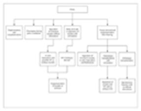
For patients with cancer, Figure 2 provides an algorithm of various possibilities. In children, it is preferable to cryopreserve ovarian tissue (described below) before treatment begins. In older patients with cancer, all cryopreservation methods are options, but freezing embryos or oocytes is preferred. Among these options, ovarian stimulation and oocyte retrieval are more reliable than the aspiration of immature oocytes for in vitro maturation; however, ovarian stimulation necessitates a delay in treatment of four to six weeks.
|
In all scenarios, the most successful approach is embryo cryopreservation. This technique affords a pregnancy rate of 20 to 30 percent per transfer of two to three embryos.12 However, this approach requires in vitro fertilization and a participating male partner, although frozen sperm from a donor may also be used. If many mature oocytes are retrieved, there is an opportunity to carry out several attempts at embryo transfer from a single cycle. Although this method is appropriate for women at great risk for ovarian failure or to extend the fertility potential of women between the ages of 38 and 40 years, it may be problematic for some women without a partner and is not applicable to children.
Oocyte cryopreservation is another potential option. Because of the fragility of the meiotic spindle and the formation of ice crystals, the success of this approach has been limited but is improving. Newer cryopreservation methods, particularly the use of vitrification (Figure 3)48,49 and intracytoplasmic sperm injection for fertilizing oocytes,48,49,50,51,52 have resulted in viable pregnancies. For this option, ovarian stimulation is used as described above in order to retrieve mature oocytes. The obstacle to the success of this approach is oocyte survival during the thawing process, which is generally about 37 percent.51 Although the pregnancy rate per cycle (after intracytoplasmic sperm injection and embryo transfer) has been reported to be as high as 22 to 25 percent,51,52 this rate is not routine, and on the basis of each embryo generated, the realistic success of this approach is only 2.2 percent,52 or a 3 percent pregnancy rate per thawed oocyte.53 Fewer than 100 births have been reported to date from oocyte cryopreservation. Because of the limited success of this technique and a lack of long-term experience with it (including data on birth outcomes), the American Society for Reproductive Medicine has recently recommended that this method not be routinely attempted and that it be carried out only under the aegis of research.54 Nevertheless, commercialization of this approach has already begun.
|
In specialized centers, aspiration of immature oocytes from fresh
tissue or follicular aspirates may be attempted so that oocytes can
then undergo in vitro maturation. The matured oocytes may be
vitrified or fertilized by intracytoplasmic sperm injection and
cryopreserved.55,56
This technique may also be used with cryopreserved tissue. These
techniques have been used in patients with cancer but mostly are
applicable to women with polycystic ovaries who are infertile.
In instances in which a large yield of harvested oocytes is desired for optimal results in cryopreservation, the use of agents to stimulate ovulation is a potential concern among patients with cancer. The aspiration of immature follicles for in vitro maturation can mitigate this concern, although this technique is not applicable to all patients or a realistic option in all centers. In addition, ovarian aspiration carries some risk of hemorrhage and infection (<1 percent). Stimulation agents should be a concern only in patients with estrogen-sensitive cancers such as breast cancer. In such cases, alternative stimulation regimens may be considered, such as the use of tamoxifen or aromatase inhibitors,57 although these regimens are less effective without added gonadotropins.
Ovarian cryopreservation is an attractive approach to the preservation of fertility and has proved successful in several animal models.58,59,60 The rationale for this approach is that primordial follicles in excised ovarian tissue may be more resistant to freezing and thawing than are mature oocytes and may be preserved without a delay in treatment. The principal obstacle to the success of this technique is poor oocyte viability. This rate is affected by the cryopreservation methods and ischemic damage, which occurs after thawing and transfer either orthotopically (to the ovarian site) or heterotopically (to another site, such as to the forearm) because of the lack of an adequate blood supply. Ischemic damage is the greater of these challenges,59,61,62 and for this reason it has been proposed to excise an intact ovary with its vascular pedicle for cryopreservation, with later vascular reanastomosis.62,63,64 However, this procedure is more surgically invasive than other approaches and has not been recommended because of the chance of spontaneous ovarian function after treatment.
Some of the debate about ovarian cryopreservation relates to its efficacy, although hundreds of samples all over the world have been frozen.65 A recent report by Donnez et al. describes the sole pregnancy occurring after orthotopic transplantation and gives encouragement to the field.66 However, in this case, the pregnancy may not have resulted from the cryopreserved tissue, since the patient had intact ovaries. This approach also remains fairly invasive, requiring two to four operations, and is somewhat inefficient.
A major concern is the short-term viability of the transplant. For an allograft to be successful, there is a small window of opportunity before the viability of the transplanted tissue is lost. Oktay et al.67,68,69 have shown that there may be only nine months to three years of endocrine function after tissue replacement. With heterotopic transplantation of autologous ovarian tissue to the forearm, Oktay and colleagues69 were able to produce a single four-cell embryo but reported no pregnancy after multiple attempts at oocyte harvesting — a finding suggesting that the site of replacement may also be an important variable. Because of the small window of opportunity, it would seem ideal to stimulate transplanted tissue in order to obtain multiple oocytes for fertilization by intracytoplasmic sperm injection. However, the viability of the tissue is such that a poor yield of oocytes is to be anticipated.70 Although intermittent ovarian failure occurs and the prognosis for spontaneous pregnancy is low in this population of patients who are at risk, Bath et al.71 recently reported a spontaneous pregnancy in a woman whose ovarian tissue had been frozen but not transplanted.
In patients with cancer, there is a risk of transmission of cancer cells from the excised ovarian tissue, although this risk appears to be low.65 Genetic problems are also possible with cryopreservation, but the risk is probably very low on the basis of several studies with oocytes.72,73 The cryopreservation of ovarian tissue should still be considered experimental54 and is not the best option for women, with or without cancer, in cases in which other options are available. Elsewhere in this issue of the Journal, Silber et al. describe a case of ovarian transplantation between monozygotic twins in which the recipient twin had unexplained premature ovarian failure.74 Although this rare case may not be directly relevant for patients with cancer, the feasibility of the microsurgical technique, in which large pieces of fresh ovarian cortex from the donor are transplanted to an atrophic ovary with documented viability, is a valuable contribution.
Table 2 lists several possible approaches to preserving fertility in women with cancer, along with evidence-based success rates. If pregnancy is desired immediately on remission, attempts may be begun soon after treatment. If this approach is considered, a more aggressive method involving an assisted reproductive technique should be used because the rate of spontaneous pregnancy is low (about 5 percent)34,35 and early menopause is to be anticipated.
|
Another potential approach is to protect primordial follicles from
chemotherapy, irradiation, or both. Pretreatment with a gonadotropin-releasing
hormone agonist or antagonist to keep the ovaries quiescent during
chemotherapy has been suggested75
but without convincing data in humans.76,77
A novel approach, which has been shown to be effective in mice, is to
suppress ovarian apoptosis by disruption of the sphingomyelinase gene
or by treatment with sphingosine-1-phosphate.78
This latter approach is untested in humans and has unknown
consequences with regard to the efficacy of cancer treatment.
Although the above techniques are all options for the treatment of patients with cancer (Figure 2), the woman who does not have cancer but wishes merely to preserve fertility presents the greatest challenge. The cryopreservation of ovarian tissue in this setting is not warranted, and the cryopreservation of oocytes has to be considered experimental.54 Because embryo cryopreservation has a reasonable success rate and has been carried out for many years, it may be a reasonable approach for appropriate candidates.
For all women, a final option to consider is oocyte donation. This technique affords the highest pregnancy rates, in the range of 40 to 50 percent per cycle.12 With this technique, age is not a factor, with pregnancy possible in women in the mid-50 years, as long as the uterus remains healthy; the latter is a potential concern in women who have undergone pelvic irradiation. Concern about a genetic risk of cancer (although rare) would be eliminated by this approach. In the future, nuclear or cytoplasmic transfer and similar techniques may be availabe for women who want to have their own genetic progeny.79 Clearly, these experimental techniques are not approved for use at the present time. Other research that may play a role in the future includes the derivation of oocytes from embryonic stem cells80 and developments in nuclear reprogramming in which germ cells can be turned into pluripotent cells.81
Conclusions
Because oocytes that are lost cannot be replaced, identifying women who are at risk for early ovarian failure, particularly among those with cancer, is important. Counseling about the various options should take into account the age of the woman and when she may wish to conceive. If pregnancy is to be delayed indefinitely, various cryopreservation options are available. Finally, if a woman is unclear about her wishes, she should be told that oocyte donation at a later time may still be an option.
Source Information
From the Department of Obstetrics and Gynecology, Columbia University College of Physicians and Surgeons and New York–Presbyterian Hospital, New York.
Address reprint requests to Dr. Lobo at the Department of Obstetrics and Gynecology, Columbia University College of Physicians and Surgeons and New York–Presbyterian Hospital, 622 W. 168th St., New York, NY 10032, or at ral35@columbia.edu.
References